Sinus Lift Surgery in CanadaLateral vs Transcrestal Techniques, Healing Biology, and CAD Cost Guide
- Sinus floor elevation is a well-established surgical technique that addresses one of the most common anatomical challenges in implant dentistry: insufficient bone height in the posterior upper jaw.
The maxillary sinuses, air-filled cavities in the cheekbones adjacent to the posterior teeth, expand downward after the upper back teeth are lost, a process called pneumatisation.









Overview
What is a sinus lift, and when is it needed?
> A sinus lift (sinus floor elevation) is a bone grafting procedure that increases the height of the upper back jaw by elevating the membrane lining the maxillary sinus and placing bone graft material in the space created. It is required when the posterior maxilla lacks sufficient bone height, typically less than 8mm, for conventional implant placement, due to natural sinus expansion following tooth loss.
Sinus floor elevation is a well-established surgical technique that addresses one of the most common anatomical challenges in implant dentistry: insufficient bone height in the posterior upper jaw. The maxillary sinuses, air-filled cavities in the cheekbones adjacent to the posterior teeth, expand downward after the upper back teeth are lost, a process called pneumatisation. This expansion reduces the available bone height between the sinus floor and the ridge crest, often to the point where standard-length implants cannot be placed without perforating into the sinus cavity.
At Stunning Dentistry, we plan all sinus lifts from CBCT-derived three-dimensional analysis of the sinus anatomy. Pre-surgical assessment includes membrane thickness measurement, identification of septa (internal bony ridges within the sinus that affect approach), and sinus status review. Cases with complex sinus anatomy, prior sinus surgery, or active sinus pathology are identified before scheduling and assessed for technique suitability.
| Approach | Residual Bone Height | Implant Timing | Complexity | Scar/Access |
|---|---|---|---|---|
| Lateral window | < 4–5mm | Staged (6 months later) | Moderate–high | Lateral sinus wall |
| Transcrestal (osteotome/piezo) | 4–8mm | Simultaneous possible | Moderate | Crestal (implant site) |
| Short implants (no lift) | 5–8mm | Immediate | Lower | No sinus entry |
Questions about this procedure?
Why the Upper Back Jaw Loses Bone
Why does the jaw shrink under the sinus after tooth loss?
> The upper posterior teeth are embedded in alveolar bone directly adjacent to the maxillary sinuses. When these teeth are removed, the alveolar bone that supported them resorbs from both the ridge crest downward and from the sinus floor upward, because the sinus expands into the vacated bone space. This bidirectional bone loss creates the sub-sinus height deficit that makes standard implant placement impossible without augmentation.
The maxillary sinuses exist in dynamic equilibrium with the surrounding alveolar bone throughout life. In dentate patients, pneumatisation is limited by the mechanical forces transmitted through the posterior teeth into the bone, forces that maintain osteoblastic activity and bone density adjacent to the sinus floor. When the posterior teeth are extracted and the alveolar bone begins to resorb, the pneumatisation process accelerates. The sinus membrane creeps downward into the resorbing space, and the remaining bone height decreases at a rate that, in some patients, exceeds 1mm per year after extraction.
At Stunning Dentistry, we advise patients with posterior maxillary tooth loss to obtain a CBCT within 6–12 months of extraction to document sub-sinus bone height before significant further pneumatisation occurs. This baseline measurement informs planning for both timing and technique selection, and may allow a less invasive transcrestal approach if acted upon before height falls below the 4mm threshold for that technique.
| Time After Extraction | Typical Sub-Sinus Height Change | Clinical Consequence |
|---|---|---|
| 0–6 months | Minimal pneumatisation | Short implants may still be feasible without lift |
| 6–24 months | Moderate height loss (1–2mm/year typical) | Transcrestal lift possible if >5mm remains |
| 2–5 years | Significant loss; <5mm common | Lateral window lift likely required |
| 5+ years | Severe pneumatisation; <3mm possible | Lateral window, staged; zygomatic as alternative |
Ready to discuss your options?
When a Sinus Lift Becomes Necessary
How do I know if I need a sinus lift?
> A CBCT scan is the definitive assessment. Sinus lifting is typically indicated when sub-sinus bone height is below 8mm (making standard 10mm+ implants impossible without sinus entry), below 5mm (requiring a staged lateral lift before implant placement), or below 4mm (requiring the most extensive augmentation before any implant is possible). The exact threshold depends on the implant system and the planned implant length.
The clinical decision for sinus floor elevation is not arbitrary, it is determined by measured bone height on CBCT and the minimum stability requirements for the chosen implant design. Most standard implants require a minimum of 8–10mm of bone for primary stability without sinus membrane violation. When sub-sinus height falls below this threshold, options are either to accept shorter implants (carrying higher long-term load risk in the posterior jaw), elevate the sinus floor to restore implant-capable bone height, or use angled implant designs that avoid the sinus entirely.
At Stunning Dentistry, we present every sinus lift patient with a treatment plan that includes the CBCT-derived measurements, the rationale for the chosen technique, the implant design planned, and the expected timeline from sinus augmentation to final restoration. No staged procedure is initiated without the patient understanding each phase and the decision points within it.
Curious about costs and timelines?
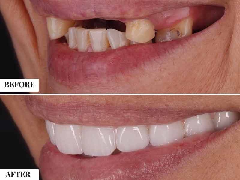
Lateral Window vs Transcrestal: Technique Comparison
What is the difference between lateral and transcrestal sinus lift?
> The lateral window sinus lift accesses the sinus through the outer cheek-side sinus wall under direct vision, allowing extensive augmentation regardless of residual bone height. The transcrestal technique approaches through the implant osteotomy from below, a less invasive approach requiring at least 4–5mm of residual bone but often allowing same-visit implant placement. Lateral lifts augment more volume and are staged; transcrestal lifts are less invasive and often simultaneous.
The lateral window sinus lift, first described by Tatum and popularised by Boyne and James, creates a bony window in the lateral sinus wall through the soft tissue via a flap reflection. The window is either in-fractured (hinged inward to become a new sinus floor) or completely removed and the membrane elevated under direct sight using curettes designed to separate the membrane from the bony sinus walls without perforation. Graft material is then packed into the sub-sinus space to the desired height. The lateral approach allows precise control of membrane elevation, augmentation volumes of 8–12mm or more, and visual management of membrane perforations when they occur.
At Stunning Dentistry, we perform both lateral and transcrestal sinus lifts and select between them based on pre-surgical CBCT anatomy. For borderline cases (5–6mm residual bone), we discuss with the patient whether simultaneous placement with a transcrestal lift or staged placement with a lateral lift better matches their timeline priorities and risk profile.
| Feature | Lateral Window | Transcrestal |
|---|---|---|
| Residual bone needed | < 4–5mm adequate | Requires ≥ 4–5mm |
| Implant timing | Staged (6 months) | Simultaneous possible |
| Augmentation volume | High (8–15mm gain) | Moderate (2–5mm gain) |
| Membrane visualisation | Direct | Indirect |
| Perforation rate | ~15–20% (manageable) | ~10–15% |
| Post-op swelling | More pronounced | Mild |
| Procedure time | 60–90 min per side | 20–40 min per site |
| Second surgery needed | Yes (implant placement) | No (if simultaneous) |
Want a personalised treatment plan?
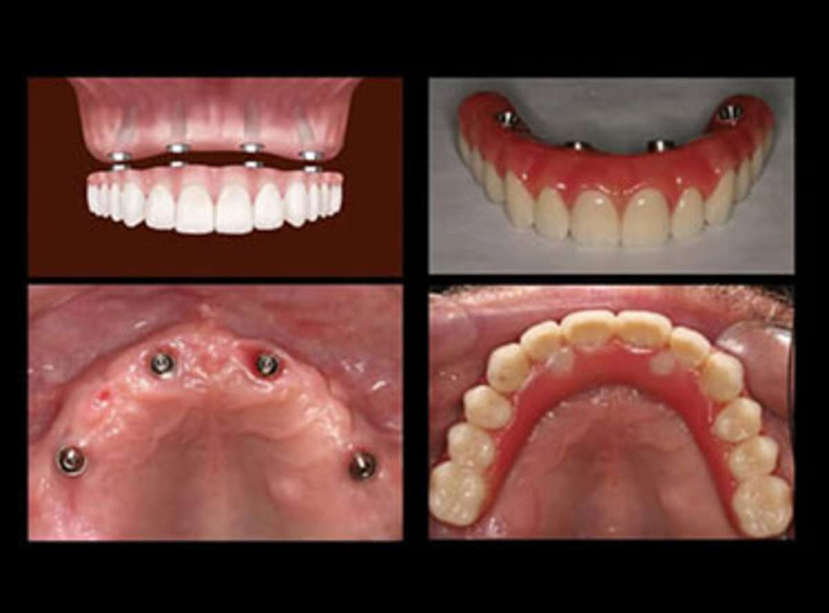
The Sinus Lift Procedure: What Actually Happens
What happens during a sinus lift?
> Under local anaesthesia, a flap is raised over the upper back jaw. For lateral lifts, a window is made in the sinus wall, the membrane is elevated, and graft material fills the space. For transcrestal lifts, instruments compress bone upward through the drill site to push the sinus membrane up before graft placement. The procedure takes 45–90 minutes per side and requires suture removal 10–14 days later.
For the lateral window technique, the surgical sequence begins with local anaesthetic infiltration and palatal nerve block, followed by full-thickness mucoperiosteal flap elevation to expose the lateral sinus wall. The window outline is scored with a bur or piezo insert, piezo is preferred as it cuts hard tissue without injuring the soft sinus membrane, and the bony window is reflected inward or removed. The Schneiderian membrane is then systematically elevated from the floor, medial, anterior, and posterior sinus walls using curved sinus curettes. The degree of elevation corresponds to the augmentation height required plus the planned implant length.
At Stunning Dentistry, we use piezo ultrasonic instruments for sinus window creation and membrane elevation in all lateral sinus lift cases, which substantially reduces Schneiderian membrane perforation risk compared to rotary bur techniques. Intraoperative membrane integrity is confirmed by Valsalva test. Perforations, when they occur, are managed immediately with collagen sponge plugs and resorbable membrane reinforcement before grafting continues, we do not abort the procedure for a manageable perforation.
| Procedural Phase | What Occurs | Duration |
|---|---|---|
| Anaesthesia | Local infiltration + posterior superior alveolar nerve block | 10–15 min |
| Flap elevation | Mucoperiosteal reflection to expose lateral sinus wall | 10–15 min |
| Window preparation (lateral) | Piezo or bur scoring and reflection | 10–15 min |
| Membrane elevation | Systematic separation from sinus walls; Valsalva check | 15–20 min |
| Graft placement | Incremental packing to planned height | 15–20 min |
| Window closure and suturing | Membrane or bone over window; tension-free flap closure | 10–15 min |
| Total per side | Single-site lateral sinus lift | 60–90 min |
| Transcrestal lift | Per-site (during implant drilling) | 20–40 min |
Questions about this procedure?
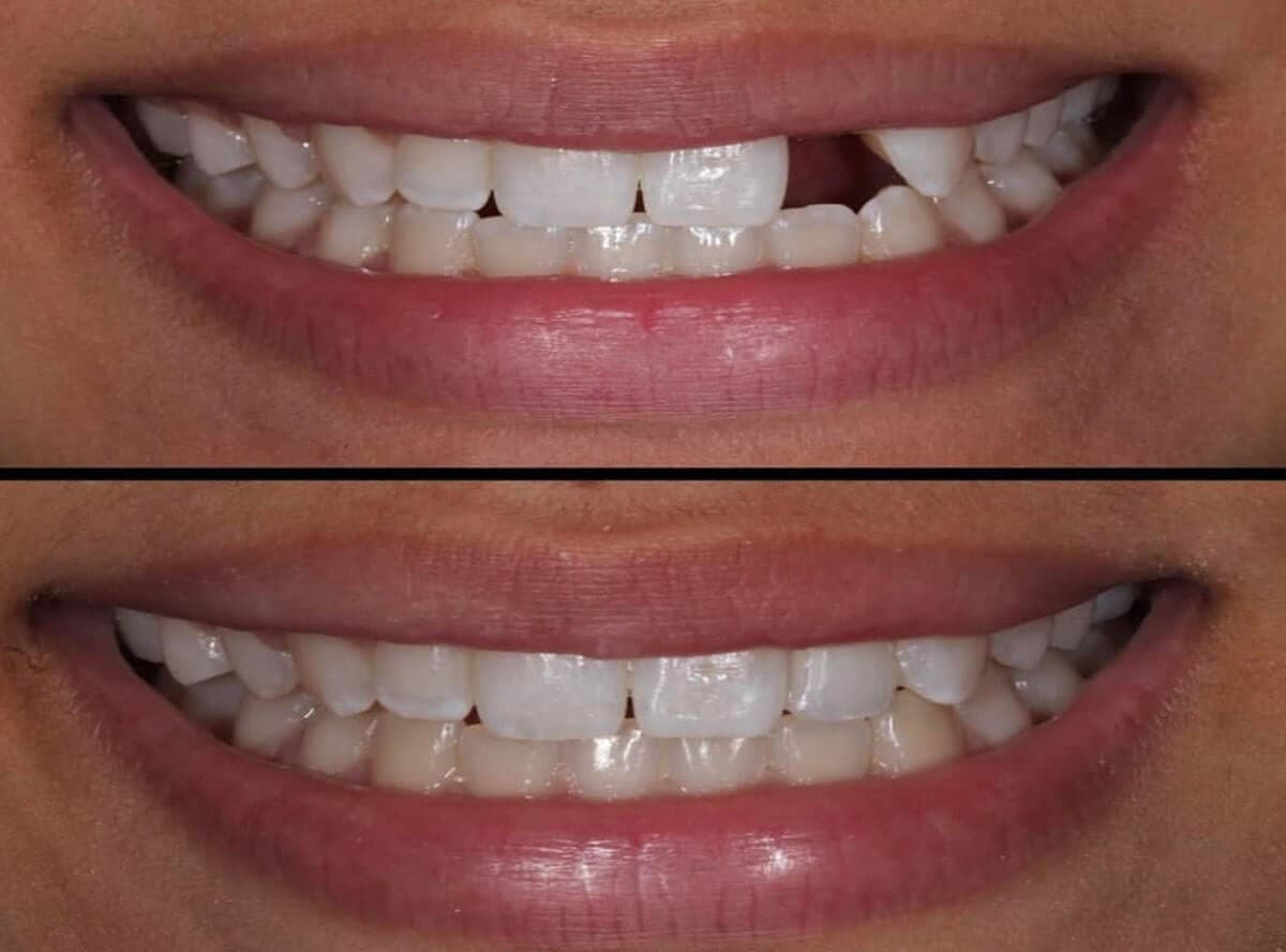
Pain and Recovery: What to Expect
How painful is sinus lift recovery?
> Most patients describe post-sinus lift discomfort as comparable to a tooth extraction, localised soreness, cheek swelling, and possible bruising for 4–7 days. Sinus congestion or mild drainage for 1–2 weeks after a lateral lift is common and expected. No nose blowing for 2 weeks is the most important post-operative restriction. Significant pain after day 3–4, facial pressure that worsens, or fever indicates a complication requiring clinical review.
Post-operative swelling after lateral sinus lifting is more pronounced than after crestal bone grafting, because the surgical access involves elevation near the infraorbital and posterior superior alveolar nerve territories, and tissue dissection is more extensive. Peri-orbital (around the eye) bruising is possible in approximately 15–20% of patients and resolves within 7–14 days. Analgesics follow the same protocol as bone grafting, ibuprofen with paracetamol, unless contraindicated. Nasal decongestants (oxymetazoline or xylometazoline, short-term) are sometimes prescribed to maintain sinus drainage and reduce post-operative sinus pressure.
The no-nose-blowing restriction is the most critical post-operative instruction for sinus lift patients. Forced nasal expiration creates sinus pressure that can dislodge the elevated membrane from its new position and displace graft material before healing stabilises the augmented space. Sneezing should be performed with an open mouth (to reduce sinus pressure) for the first 2 weeks. Swimming and diving are avoided for 4 weeks; air travel is permitted after 72 hours for most patients, with decongestant coverage.
You should plan for 5–7 days of reduced activity and social restriction for swelling management after a lateral sinus lift. The transcrestal approach causes significantly less post-operative swelling, most patients report only mild soreness for 2–3 days. During the 6-month maturation phase after a lateral lift, the only ongoing restriction is avoidance of posterior denture pressure over the graft site; patients may return to normal diet within 3–4 weeks once the surgical wound heals.
At Stunning Dentistry, our post-operative protocol for sinus lift patients includes a written post-op guide with instructions specific to sinus procedures (nose restriction, decongestant use, activity), a 48-hour phone check-in from the clinical coordinator, and a 2-week clinical review. Remote patients returning to Canada before the 2-week review receive written guidance for local GP assessment and are in contact with our team remotely for the first post-operative month.
| Recovery Phase | Expected Experience | Key Restrictions |
|---|---|---|
| Day 1–3 | Cheek swelling, soreness, possible periorbital bruising | No nose blowing; soft foods; decongestant |
| Day 4–10 | Swelling decreasing; nasal congestion settling | Mouth-open sneezing; no strenuous activity |
| Day 10–14 | Switure removal; site healing well | Soft diet continuing |
| Month 1–6 | Asymptomatic maturation phase | No removable denture pressure over graft |
| Month 6 | CBCT confirms sub-sinus bone height | Implant placement appointment scheduled |
Ready to discuss your options?
Risk Transparency
What are the risks of a sinus lift?
> Schneiderian membrane perforation during elevation is the most common intraoperative complication, occurring in approximately 10–30% of lateral sinus lifts depending on membrane thickness and technique. Most perforations are manageable and do not prevent the procedure from continuing. Post-operative sinusitis occurs in 2–5% of cases. Graft migration, infection, and implant failure in grafted bone are less common but clinically significant risks that affect a minority of patients.
The Schneiderian membrane (the mucosal lining of the sinus) is approximately 0.3–0.8mm thick and adheres to the bony sinus walls with varying degrees of resistance. Thin membranes, common in patients with prior sinusitis, smoking history, or anatomical variation, are more susceptible to tearing during elevation. Membrane perforations are classified by size: small (< 5mm) are managed with resorbable collagen membrane reinforcement and the procedure continues; large perforations (> 10mm) may require aborting the lateral lift and rescheduling after 2–3 months of membrane healing. The use of piezo ultrasonic instruments significantly reduces perforation frequency compared to rotary burs.
At Stunning Dentistry, all pre-surgical sinus lift CBCT assessments include a formal sinus pathology screen. Any finding of membrane thickening greater than 2mm, mucosal retention cysts, mucous plugging, or septal deviation is documented and discussed with the patient. Where the clinical picture suggests active sinus disease, we refer to ENT for clearance and possible pre-treatment before scheduling the sinus lift.
| Complication | Frequency | Management | Effect on Outcome |
|---|---|---|---|
| Membrane perforation (small, < 5mm) | 10–20% | Collagen reinforcement; proceed | Usually no impact |
| Membrane perforation (large, > 10mm) | 2–5% | Abort and reschedule; 2–3 months healing | Delays by 3 months |
| Post-operative sinusitis | 2–5% | Antibiotics, decongestants; ENT if severe | Usually resolved; rare graft removal |
| Graft migration into sinus | 1–3% | Endoscopic removal if symptomatic | May compromise graft volume |
| Inferior alveolar nerve involvement | Not applicable (maxillary procedure) | N/A | N/A |
| Implant failure in grafted sinus | 3–8% at 5 years | Re-implantation after healing | Extends timeline |
Curious about costs and timelines?
Clinical Success Determinants
Lateral sinus lift survival rates for implants placed in grafted sinus bone range from 90–95% at 5 years in systematic reviews, broadly comparable to implants placed in native bone at the same site. This outcome is technique- and material-dependent: studies using xenograft (Bio-Oss) and piezo technique report the highest survival rates. The long-term maintenance of grafted sub-sinus volume, with no significant height loss observed at 5–10 year follow-up, is one of the most reproducible outcomes in implant augmentation surgery.
| Success Determinant | Why It Matters | What to Verify |
|---|---|---|
| Pre-surgical sinus patency | Obstructed sinus drainage increases post-op sinusitis risk | CBCT membrane check; ENT clearance if thickening >2mm |
| Membrane integrity at elevation | Unmanaged large perforations compromise graft space | Ask about perforation frequency and management protocol |
| Piezo vs rotary instrument use | Piezo reduces membrane perforation rate | Confirm piezo is used for window creation and elevation |
| Graft material selection | Slowly resorbing xenograft (Bio-Oss) maintains volume long-term | Confirm material used and its clinical evidence base |
| Primary wound closure | Exposed graft or membrane invites infection | Ask about flap design and suturing technique |
| No nose blowing compliance | Critical in first 2 weeks to protect membrane position | Patient must understand this restriction clearly |
| Non-smoking status | Impairs vascular ingrowth and graft integration | Cessation for full maturation period |
| Adequate maturation time | Premature implant placement risks failure | Implant only after CBCT confirms sub-sinus bone density |
| No active sinus pathology | Sinusitis creates bacterial contamination of graft | Pre-op ENT evaluation for high-risk patients |
Want a personalised treatment plan?
Healing Timeline
Transcrestal lift with simultaneous implant: Crown placement approximately 8–10 months from initial surgery when implant and simultaneous lift are performed together.
Bilateral simultaneous lifts: Both sides can be performed at a single surgical visit; timeline is the same as unilateral.
| Phase | Timeline | Biological Activity | Clinical Marker |
|---|---|---|---|
| Clot and membrane stabilisation | Week 1–2 | Fibrin matrix organises; membrane seals over graft | Sutures removed; no symptoms |
| Early graft vascularisation | Month 1–2 | Blood vessels penetrate graft from sinus floor and window margins | Asymptomatic; soft diet |
| Active bone formation | Month 2–5 | Osteoblast colonisation of scaffold; mineralisation begins | Asymptomatic maturation |
| Sub-sinus bone maturation | Month 5–6 | New bone density increases; scaffold integrates | CBCT confirms height and density |
| Implant placement | Month 6–9 | After CBCT confirmation | Implant surgery and primary healing (4 months) |
| Final restoration | Month 10–14 from lift | Implant osseointegrated | Crown placement |
Questions about this procedure?
When Sinus Lifting Is Not Recommended
Who should not have a sinus lift?
> Active maxillary sinusitis is an absolute contraindication, the graft would be placed in a bacterially contaminated space. Prior extensive sinus surgery (radical antrostomy, total Caldwell-Luc) that has disrupted the sinus architecture or eliminated the Schneiderian membrane makes standard lifting technically impossible. Severe immunosuppression, IV bisphosphonate use, and head/neck radiation are the same systemic contraindications that apply to all elective bone grafting. Relative contraindications include poorly controlled allergic rhinitis, chronic rhinosinusitis, and smoking.
Chronic maxillary sinusitis with active bacterial or fungal infection requires ENT evaluation and treatment before any sinus lift is considered. The CBCT findings in such cases typically show complete or partial sinus opacification, thickened mucosa, or air-fluid levels. Introducing graft material into an infected sinus reliably produces graft contamination, failure, and possible spread of infection. ENT clearance with confirmed sinus health on repeat CBCT is required before surgery is scheduled.
At Stunning Dentistry, we use CBCT-based sinus pathology scoring before all sinus lift cases. Findings that place a patient at elevated risk are discussed openly, and cases requiring ENT evaluation are referred before any surgical date is given. Patients who need pre-treatment before sinus lifting receive a written staged plan explaining what must occur, in what sequence, and what the timeline implications are.
| Contraindication | Type | Clinical Action |
|---|---|---|
| Active maxillary sinusitis | Absolute | ENT treatment required before any planning |
| Radical prior sinus surgery (membrane absent) | Absolute | Zygomatic implants as alternative |
| IV bisphosphonate therapy | Absolute | Oncology consultation required |
| Head/neck radiation > 50 Gy | Absolute | High MRONJ risk; specialist evaluation |
| Membrane thickening > 2mm on CBCT | Relative | ENT clearance; pre-treat if symptomatic |
| Nasal polyps | Relative | ENT polypectomy before lifting |
| Deviated septum impeding drainage | Relative | ENT septoplasty may be indicated pre-lift |
| Smoking | Relative | Cessation required for maturation period |
| Poorly controlled diabetes | Relative | Optimise HbA1c < 7.5% before surgery |
Ready to discuss your options?
Alternative Options
When a sinus lift is contraindicated or the patient declines staged augmentation, zygomatic implants offer the most direct alternative for the posterior maxilla. Zygomatic implants (40–52mm long) bypass the maxillary sinus entirely, anchoring in the dense cortical bone of the zygomatic arch. They require specific surgical training and are indicated for severely resorbed maxillae or cases where the sinus cannot be augmented. Published 10-year survival rates of 95%+ in specialist centres make them a reliable long-term option.
At Stunning Dentistry, our surgical team performs both sinus augmentation and zygomatic implant procedures. Patients presenting with severe posterior maxillary bone loss are evaluated for both options, with a written comparison of the clinical rationale, timeline, and outcome expectations for each path before a decision is made.
Curious about costs and timelines?
Cost Logic: CAD Comparison
Materials note: Stunning Dentistry uses Geistlich Bio-Oss xenograft and Geistlich Bio-Gide collagen membranes, the same materials used in Canadian oral surgery practices. Piezo instruments used for window preparation and membrane elevation match the standard of specialist surgical centres. The cost differential reflects Canadian overhead, specialist fee schedules, and surgical facility charges, not material or technical difference.
| Procedure | Stunning Dentistry (India), CAD | Canadian Oral Surgeon, CAD | Difference |
|---|---|---|---|
| Transcrestal sinus lift (1 site) | $600 – $1,000 | $1,200 – $2,500 | ~$600 – $1,500 saved |
| Lateral window sinus lift (1 side) | $1,500 – $2,500 | $3,000 – $6,000 | ~$1,500 – $3,500 saved |
| Bilateral lateral sinus lift | $2,800 – $4,500 | $6,000 – $12,000 | ~$3,200 – $7,500 saved |
| Sinus lift + implant (staged, 1 side) | $3,500 – $5,500 | $7,000 – $14,000 | ~$3,500 – $8,500 saved |
| Sinus lift + implant (simultaneous) | $2,500 – $4,000 | $5,000 – $10,000 | ~$2,500 – $6,000 saved |
Want a personalised treatment plan?
Treatment Comparison Matrix
| Factor | Lateral Sinus Lift | Transcrestal Lift | Short Implants | Zygomatic Implants |
|---|---|---|---|---|
| Minimum residual bone | None required (< 1mm feasible) | 4–5mm | 5–8mm | Not dependent on alveolar bone |
| Implant timing | Staged (6 months) | Simultaneous | Immediate | Immediate |
| Augmentation volume | High (up to 15mm) | Moderate (2–5mm) | None | None |
| Complication profile | Membrane perf, sinusitis | Membrane perf (less frequent) | Higher fracture risk | Surgical complexity, sinus adjacency |
| Total treatment time to crown | 14–18 months | 8–10 months | 4–6 months | 4–6 months |
| Long-term sinus bone stability | Excellent (xenograft scaffold) | Good | N/A | N/A |
| Cost (CAD, SD) | $1,500–$2,500 | $600–$1,000 | Lower (no lift) | Higher (complex) |
| Best for | < 4mm residual bone | 4–8mm residual bone | Borderline height with stable width | Severe resorption, failed grafts |
Questions about this procedure?
Post-Procedure Biological Reality
The new bone formed beneath an elevated sinus membrane develops from multiple biological sources: osteoblasts migrating from the cut sinus floor edges, mesenchymal stem cells mobilised by the surgical stimulus, and osteoprogenitor cells from the periosteum at the lateral window margins. This multi-directional bone formation characteristic of sinus augmentation is one reason the sinus lift has become one of the most predictable procedures in implant augmentation surgery, bone forms from the walls inward, not just from a single base.
At Stunning Dentistry, CBCT at 6 months post-lateral-lift documents the sub-sinus bone height achieved, the density (measured in Hounsfield units at the planned implant axis), and any radiographic abnormalities within the sinus. Implant placement proceeds only when the three-dimensional analysis confirms adequate volume and density for the planned implant design and length. This measurement-based confirmation, rather than elapsed time alone, is the trigger for proceeding to the implant phase.
Ready to discuss your options?
Common Mistakes
What causes sinus lift failure?
> The most common causes are: operating in an undiagnosed or inadequately cleared sinus pathology; proceeding with a transcrestal approach when sub-sinus bone is too thin for primary stability; unmanaged membrane perforation that contaminates the graft; patient non-compliance with the no-nose-blowing restriction; and premature implant placement before graft maturation is confirmed by CBCT.
Performing a sinus lift without pre-operative CBCT is the most foundational mistake in this procedure. Panoramic X-ray alone is insufficient to assess membrane thickness, sinus septum anatomy, the presence of retention cysts, or the three-dimensional relationship of the sinus floor to the planned implant axis. Surgeons who plan sinus augmentations from panoramic radiographs alone are operating with an incomplete picture of sinus anatomy that increases the rate of intraoperative complications.
At Stunning Dentistry, no sinus lift is planned without CBCT-derived measurements. All technique selections are documented with the specific anatomical rationale, and patients receive the CBCT images and the report as part of their treatment file.
Curious about costs and timelines?
Myth Deconstruction
Myth: "A sinus lift is a high-risk major surgery."
The majority of sinus lifts use xenograft (bovine bone mineral, Bio-Oss), no second surgical site. Autogenous bone is used in some complex cases requiring faster bone formation in the sinus cavity, but it represents a minority of clinical cases. The evidence for xenograft in sinus augmentation is extensive and spans more than 25 years of clinical follow-up.
Want a personalised treatment plan?
People Also Ask
How long does it take to recover from a sinus lift?
Yes. If a sinus lift produces insufficient bone volume or the graft fails, re-augmentation after a healing period of 3–6 months is a recognised option. Success rates for secondary sinus augmentation are modestly lower than for primary procedures but remain clinically significant. Zygomatic implants are an alternative if sinus augmentation cannot be successfully achieved.
Questions about this procedure?
Ask Your Doctor
- What is the sub-sinus bone height at my implant sites, measured from the CBCT scan?
- Will you use piezo instruments or a rotary bur for window preparation and membrane elevation?
- What graft material will you use, and why is it appropriate for my case?
- Do you perform lateral and transcrestal sinus lifts, and which technique is indicated for my anatomy?
- What is your rate of membrane perforations, and how do you manage them intraoperatively?
- Is there any sinus pathology on my CBCT that requires ENT evaluation before you will proceed?
- When and how will you confirm that the graft has matured sufficiently before placing the implant?
- What are the restrictions on nose blowing, exercise, and air travel after surgery?
Ready to discuss your options?
Curious about costs and timelines?
For Canadian Patients
Pre-Travel Checklist for Canadian Patients Considering Sinus Lift in India
"addressCountry": "IN"
| Step | Action | When |
|---|---|---|
| 1 | Obtain CBCT scan at Canadian imaging centre (sub-sinus height must be measured and documented in DICOM) | Before booking travel |
| 2 | Send DICOM files to Stunning Dentistry for sinus anatomy analysis and technique planning | 4–6 weeks before travel |
| 3 | Disclose sinus history (sinusitis, prior sinus surgery, nasal polyps, allergic rhinitis) in intake form | Pre-travel |
| 4 | Obtain ENT clearance if any sinus pathology history | Pre-travel |
| 5 | Arrange to stay near the clinic for minimum 5–7 days post-surgery for monitoring and suture review | Pre-travel |
| 6 | Arrange local ENT or GP in Canada for post-operative review if returning before 2 weeks | Pre-travel |
| 7 | Arrange local imaging centre for 6-month post-lift CBCT | At 4–5 months post-lift |
| 8 | Book return visit for implant placement after CBCT confirms maturation | At 6–9 months |
Want a personalised treatment plan?
Clinical References
1. Boyne PJ, James RA. "Grafting of the maxillary sinus floor with autogenous marrow and bone." *Journal of Oral Surgery* 1980;38(8):613–616.
*Diagnosis precedes decision.*
Questions about this procedure?
Specialist-only treatment planning
- Remote file review before travel
- Evidence-led treatment checkpoints
No waiting list for eligible cases
- Remote file review before travel
- Evidence-led treatment checkpoints
Trip coordinated with care timeline
- Remote file review before travel
- Evidence-led treatment checkpoints
Our Partners











Why Us
Frequently Asked Questions
Can I have a sinus lift if I have had prior sinus surgery?
Prior sinus surgery does not necessarily preclude a sinus lift, but it requires individualised evaluation. Conservative procedures such as FESS (functional endoscopic sinus surgery) or antrostomy that have preserved the sinus architecture and the Schneiderian membrane may not compromise augmentation. Radical procedures that have removed significant sinus mucosa or architecture require specialist assessment before planning.
Can a sinus lift be done at the same time as tooth extraction?
Immediate sinus lifting at the time of posterior tooth extraction (simultaneous extraction and sinus grafting) is a described approach for selected cases. It requires that the extraction socket be free of active infection, that the residual bone permits the planned technique, and that the sinus membrane is intact at the extraction site. This approach compresses the treatment timeline but is not universally applicable.
What type of sedation is available for sinus lift surgery?
Most sinus lift procedures are performed under local anaesthesia, the patient is awake but the surgical site is fully anaesthetised. Intravenous conscious sedation (midazolam, propofol) or oral sedation is available for anxious patients at Stunning Dentistry. General anaesthesia is not routinely used for sinus lift procedures and is unnecessary for the large majority of patients.
Will I need antibiotics before and after the sinus lift?
Antibiotic prophylaxis is standard practice for sinus lift procedures, typically amoxicillin 2g 1 hour pre-operatively (or clindamycin 600mg for penicillin-allergic patients) and a post-operative course of 5–7 days. Post-operative nasal decongestants are often prescribed to maintain sinus drainage patency during the early healing phase.
See your new smile instantly!
This tool will help you understand potential structural and aesthetic changes before finalizing treatment decisions.